Quantitative imaging · Host biology
HostBio / Comp-MRF
Treating routine clinical CT as a quantitative measurement of host tissue state, then asking whether the inferred phenotype survives contact with biology outside the image.
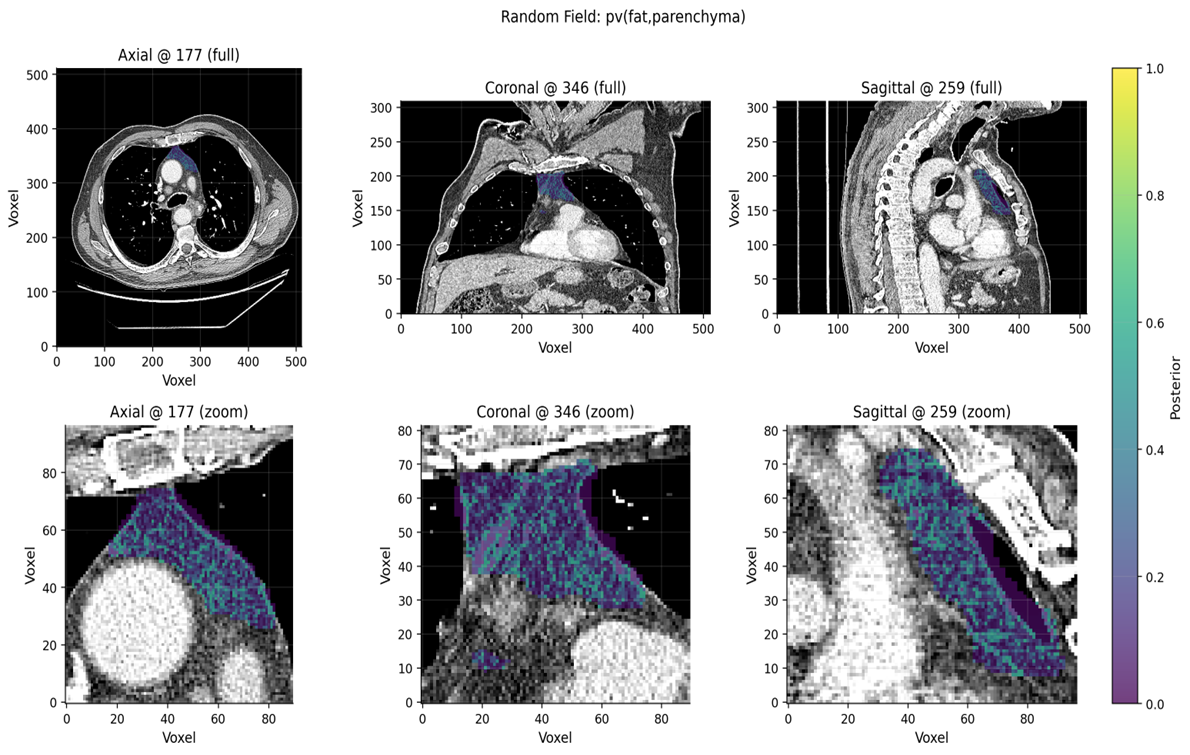
Clinical CT is acquired repeatedly and at enormous scale, but scanner settings, contrast, anatomy, and partial-volume mixing make raw intensity difficult to interpret biologically. A thresholded organ volume can therefore miss the part of the signal that matters most: tissue composition.
I developed HostBio around a different question: can routine imaging be turned into a stable, interpretable measurement of host tissue state that can then be challenged with independent biological data?
The original insight came from recognizing a structural analogy with deconvolution problems in spatial transcriptomics: a measured unit can contain mixtures of underlying biological components. The analogy is useful, but the physics are different. CT requires a model of partial-volume mixing, acquisition variability, contrast, and spatial dependence rather than a transcript-count model.
Comp-MRF therefore estimates tissue-composition posteriors using scan-adaptive mixture modeling, spatial regularization, nuisance-component handling, and within-scan reference tissues. The output remains interpretable at the level of biological composition rather than becoming an opaque image embedding.
I validate HostBio-derived thymic phenotypes against measurements that do not come from CT. Across 1,078 patients in four independent NSCLC cohorts, including randomized RTOG-0617, the analyses integrate immune profiling, targeted tumor and cfDNA sequencing, treatment exposure, toxicity, response, and survival.
These studies identify treatment-context-dependent associations with outcome and toxicity, and link lower thymic reserve to persistent suppressive or exhaustion-associated peripheral immune programs and impaired treatment-emergent CD8+ proliferation. The point is not that CT substitutes for immunology. It is that imaging can provide a scalable host phenotype whose biological meaning can be tested directly.
The same framework is being extended beyond thymus to coordinated phenotyping of skeletal muscle, adipose tissue, liver, adrenal, thyroid, and other host compartments. In resectable NSCLC, ongoing work integrates thymic activity and myosteatosis with PD-L1, tumor genomics, chromosomal instability, toxicity, and pathologic response to study host immunometabolic state.
This broader program also includes deep-learning segmentation, pretrained CT risk models, serial cone-beam CT, and longitudinal imaging. The common objective is not simply better image analysis. It is to define quantitative measurements that can support biological and clinical inference.
Automated segmentation makes cohort-scale analysis possible, but HostBio is aimed at a different problem: extracting quantitative host phenotypes from routine clinical imaging and determining when those phenotypes represent meaningful immune and tissue biology.
That distinction is central to how I approach computational biology. A model is most useful when its representation has a clear measurement interpretation, its assumptions and uncertainty are visible, and its outputs can be challenged with orthogonal data.
Selected outputs
Related work
Thymic Composition Predicts Radiation Pneumonitis in Locally Advanced NSCLC
CT-Derived Thymic Involution Is Associated with a Peripheral CD8+ Inhibitory-Receptor Phenotype and Inferior Survival in Unresected Locally Advanced NSCLC
Thymus Composition, Disease Control, and Toxicity in Locally Advanced Lung Cancer
CT-Derived Host Immuno-Metabolic Phenotyping Predicts Response to Neoadjuvant Chemoimmunotherapy in Resectable Locally Advanced NSCLC